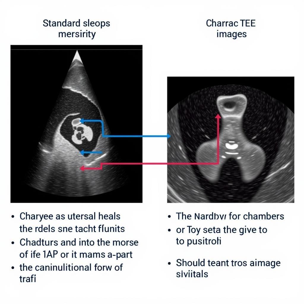
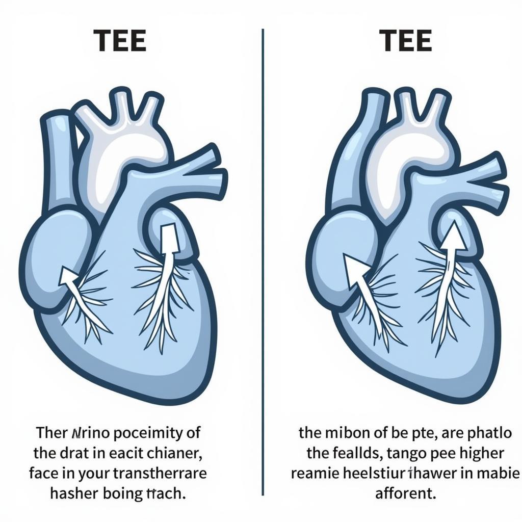
Ase Transesophageal Echocardiography (TEE) is a specialized type of echocardiogram that provides detailed images of the heart. Unlike a standard echocardiogram, where the ultrasound probe is placed on the chest, a TEE involves inserting a small probe into the esophagus, the tube that connects the mouth to the stomach. This closer proximity to the heart allows for clearer, more precise images, particularly of the heart’s valves, chambers, and surrounding structures. TEE plays a crucial role in diagnosing various heart conditions.
It is important to note that “ASE” refers to the American Society of Echocardiography, a leading organization that develops guidelines and standards for echocardiography practices. Understanding these guidelines, such as those found in the ASE Guidelines for Echocardiography, is essential for performing and interpreting TEE accurately. These guidelines ensure that healthcare professionals adhere to best practices, resulting in optimal patient care and accurate diagnoses. This article aims to delve deeper into the procedure, benefits, risks, and applications of ASE transesophageal echocardiography.
What is ASE Transesophageal Echocardiography (TEE)?
TEE is an advanced imaging technique providing highly detailed images of the heart’s structures. It utilizes high-frequency sound waves emitted from a probe inserted into the esophagus, which then bounce back and are interpreted by a computer to create moving images. This method provides a clearer view compared to traditional transthoracic echocardiography because the ultrasound waves don’t have to travel through skin, bone, and lung tissue.
A physician specializing in cardiac imaging or an anesthesiologist usually performs the procedure. Before the procedure, the throat is numbed, and a sedative is often administered to ensure patient comfort. The small, flexible probe is then gently guided down the esophagus. The procedure typically takes about 30 to 60 minutes.
Benefits of ASE Transesophageal Echocardiography
TEE offers numerous advantages, making it an invaluable tool in cardiology. Its unique positioning allows for a closer look at heart structures, enabling the detection of subtle abnormalities that might be missed with other imaging techniques. It’s particularly useful for assessing heart valves, identifying blood clots, and evaluating the aorta, the main artery leaving the heart. TEE also plays a vital role in guiding complex cardiac procedures.
For instance, during heart valve surgery, TEE can provide real-time images, guiding surgeons with precision. The ASE Comprehensive TEE Echocardiography guidelines provide valuable information regarding the specific applications and protocols for TEE use in such scenarios. This ensures the procedure is carried out with utmost accuracy and safety, ultimately contributing to better patient outcomes.
 Benefits of ASE TEE in Cardiology
Risks and Considerations of TEE
While generally safe, TEE, like any medical procedure, carries potential risks. These can include minor discomfort in the throat, temporary hoarseness, or, rarely, more serious complications like esophageal perforation. However, these risks are minimized with proper preparation and skilled execution of the procedure. Following the ASE guidelines for echocardiography, including those specific to TEE, is crucial for minimizing potential complications. For further information on specific guidelines related to infective endocarditis, you can refer to the ASE Guidelines Infective Endocarditis. You can also find related information in ASE Guidelines Endocarditis. Another useful resource is the ASE Guidelines for Echocardiography.
When is ASE Transesophageal Echocardiography Used?
TEE is often used in situations where a standard echocardiogram doesn’t provide sufficient detail or when a clearer view of specific heart structures is needed. Some common scenarios include evaluating heart valve disease, detecting blood clots, diagnosing congenital heart defects, and guiding complex heart surgeries. Additionally, ASE Comprehensive Echocardiography PDF can provide more in-depth information regarding its various applications. Another valuable resource for in-depth information is ASE Comprehensive TEE Echocardiography.
FAQ
- Is TEE painful? While there might be some mild discomfort, the throat is numbed before the procedure, and sedation is often used to ensure patient comfort.
- How long does the procedure take? Typically, a TEE procedure lasts between 30 and 60 minutes.
- What are the possible complications of TEE? While rare, potential complications include minor throat discomfort, hoarseness, or, in very rare cases, esophageal perforation.
- How do I prepare for a TEE? Your doctor will provide specific instructions, but generally, you’ll need to fast for several hours before the procedure.
- When will I get the results? Your doctor will discuss the results with you after the procedure or at a follow-up appointment.
- Is TEE covered by insurance? Coverage varies depending on your insurance plan and the reason for the TEE. It’s best to contact your insurance provider for specific information.
- What is the difference between TEE and a regular echocardiogram? TEE provides a closer, more detailed view of the heart, especially the valves and chambers, due to the probe’s placement in the esophagus.
 TEE vs. Standard Echocardiography
Conclusion
ASE transesophageal echocardiography (TEE) is a valuable diagnostic tool that provides critical information about the heart’s structure and function. While carrying potential risks, these are minimal when performed by experienced professionals adhering to ASE guidelines. TEE plays a pivotal role in diagnosing and managing various heart conditions, contributing significantly to improved patient care and outcomes.
Common Scenarios and Questions
Patients often have questions about the discomfort and recovery process after TEE. Most experience mild throat soreness, which typically resolves within a day or two. It’s crucial to follow your doctor’s post-procedure instructions, including dietary restrictions.
Further Exploration
For more information, you might find articles on related topics like atrial fibrillation, heart valve disease, and congenital heart defects helpful. Exploring these resources can provide a broader understanding of the conditions often investigated using TEE.
For further assistance, please contact us at Phone Number: 0369020373, Email: [email protected] or visit our address: Thôn Ngọc Liễn, Hiệp Hòa, Bắc Giang, Việt Nam. We have a 24/7 customer support team.

