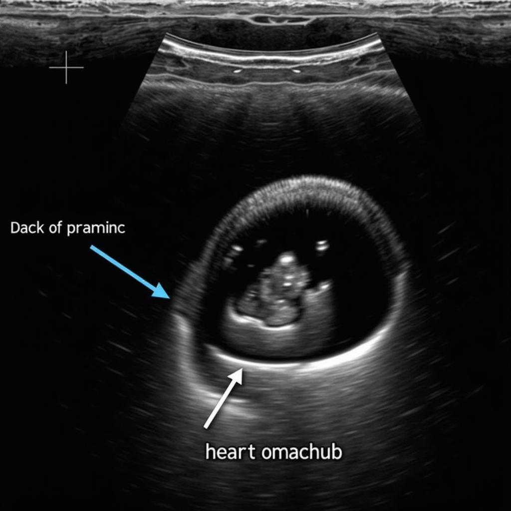
Pericardial effusion, the accumulation of fluid within the pericardium (the sac surrounding the heart), can be a serious condition. When evaluated with advanced echocardiography techniques, as often recommended by the American Society of Echocardiography (ASE), the diagnosis and management can be significantly improved. Understanding Ase Pericardial Effusion guidelines is crucial for healthcare professionals in Southeast Asia and worldwide.
Pericardial effusions can range from small, asymptomatic accumulations to large effusions causing significant hemodynamic compromise, a condition known as cardiac tamponade. The ASE provides comprehensive guidelines for evaluating pericardial effusions, encompassing both the diagnostic process and recommendations for treatment. These guidelines leverage the power of echocardiography, a cornerstone in cardiovascular imaging, to accurately assess the effusion size, location, and impact on cardiac function. The accurate assessment of ASE pericardial effusion is crucial for effective management.
Echocardiography’s Role in ASE Pericardial Effusion Assessment
Echocardiography is the primary imaging modality for diagnosing and evaluating pericardial effusions. ASE guidelines detail the specific echocardiographic views and measurements needed to quantify the effusion and assess its impact on the heart. This information is vital for determining the appropriate course of action, which can range from watchful waiting for small, asymptomatic effusions to urgent pericardiocentesis for large effusions causing tamponade. You can learn more about pericardial disease from our dedicated ASE pericardial disease resource.
Utilizing ASE Comprehensive Echocardiography for Accurate Diagnosis
The ASE promotes comprehensive echocardiography, utilizing various modalities like M-mode, 2D, and Doppler, to gain a complete understanding of the pericardial effusion. M-mode can demonstrate diastolic collapse of the right atrium and ventricle, a sign suggestive of tamponade. 2D echocardiography allows for visualization of the effusion’s location and extent, while Doppler helps assess the impact on blood flow. For more in-depth information on advanced echocardiography techniques, you can explore our resource on ASE comprehensive echocardiography.
 Echocardiography Image of Pericardial Effusion
Furthermore, the ASE emphasizes the importance of integrating clinical findings with echocardiographic data. Symptoms such as chest pain, shortness of breath, and lightheadedness, along with physical exam findings like muffled heart sounds and jugular venous distension, can raise suspicion for pericardial effusion and prompt further investigation with echocardiography.
Managing ASE Pericardial Effusion: Guidelines and Recommendations
The ASE provides detailed guidelines for the management of pericardial effusions, which vary depending on the size of the effusion, the presence of hemodynamic compromise, and the underlying cause. Small, asymptomatic effusions may simply require observation and repeat echocardiography to monitor for changes. However, larger effusions, especially those causing symptoms or signs of tamponade, require more aggressive intervention.
ASE Tamponade Guidelines: Prompt Recognition and Treatment
Cardiac tamponade is a life-threatening complication of pericardial effusion. The ASE offers specific guidelines for recognizing and managing tamponade, emphasizing the importance of prompt diagnosis and treatment. Echocardiography plays a crucial role in identifying the characteristic signs of tamponade, such as diastolic collapse of the right-sided heart chambers and exaggerated respiratory variation in mitral and tricuspid valve inflow velocities. More information on this critical condition can be found in our ASE tamponade resource.
For patients with tamponade, immediate pericardiocentesis (drainage of the pericardial fluid) is often necessary to relieve the pressure on the heart and restore normal cardiac function. The ASE guidelines provide recommendations on the techniques and precautions for performing pericardiocentesis safely and effectively. For a comprehensive understanding of managing pericardial diseases, you can also visit our ASE guidelines pericardial disease page.
Conclusion
ASE pericardial effusion guidelines are invaluable for healthcare professionals involved in the diagnosis and management of this condition. By utilizing comprehensive echocardiography and following the ASE recommendations, clinicians can accurately assess the effusion, identify potential complications like tamponade, and implement appropriate treatment strategies, ultimately improving patient outcomes. Understanding and adhering to these guidelines are crucial for providing optimal care for patients with pericardial effusion in Southeast Asia and globally.
FAQ
- What is ASE pericardial effusion?
- What are the symptoms of pericardial effusion?
- How is pericardial effusion diagnosed?
- What are the treatment options for pericardial effusion?
- What is cardiac tamponade, and how is it related to pericardial effusion?
- What are the long-term implications of having a pericardial effusion?
- Where can I find more information on ASE guidelines for pericardial disease?
Do you have other questions about pericardial effusion, echocardiography or related cardiovascular issues? Explore our ASE cardio oncology resources for further insights.
Need assistance? Contact us 24/7: Phone: 0369020373, Email: [email protected] or visit us at: Thon Ngoc Lien, Hiep Hoa, Bac Giang, Vietnam.

