The Ase Velocity Map, a crucial tool in echocardiography, allows healthcare professionals to visualize and quantify blood flow within the heart. It provides valuable insights into cardiac function and helps diagnose various cardiovascular conditions. This article delves into the significance of ASE velocity maps, their applications, and how they contribute to enhanced patient care within the ASEAN region and globally.
What is an ASE Velocity Map?
ASE velocity maps, guided by the American Society of Echocardiography (ASE), are graphical representations of blood flow velocity within the heart chambers and vessels. These maps use color-coding to depict the speed and direction of blood flow, providing a dynamic view of cardiac hemodynamics. By analyzing these maps, clinicians can assess valve function, detect abnormal flow patterns, and evaluate the overall performance of the heart.
After the initial assessment, further investigations might be needed. ase echo guidelines diastolic dysfunction provides valuable information on assessing diastolic dysfunction.
How Does it Work?
ASE velocity maps are generated using Doppler echocardiography, a technique that utilizes ultrasound waves to measure blood flow velocity. The Doppler effect, a change in the frequency of sound waves as they reflect off moving objects, is applied to calculate blood flow speed and direction. This data is then processed and displayed as a color-coded map superimposed on a 2D echocardiographic image.
Interpreting the Colors
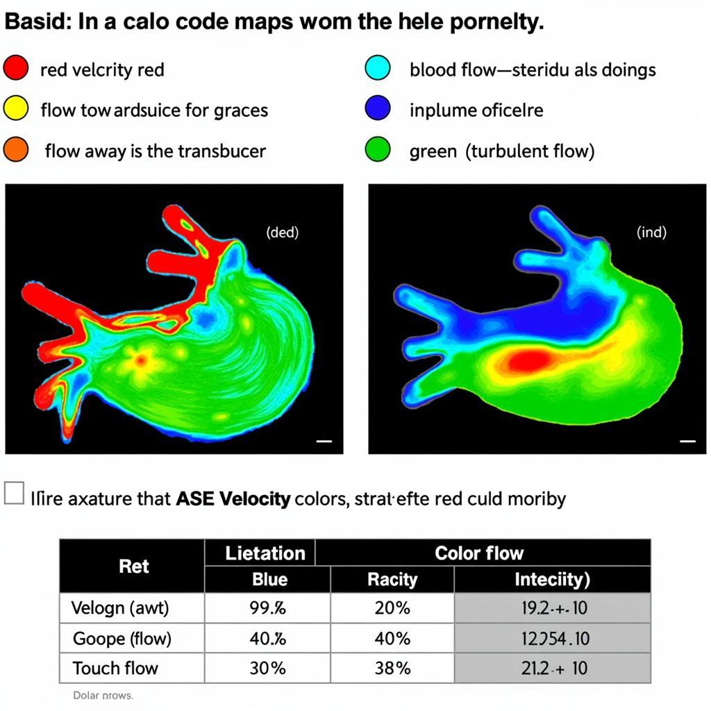
The color scheme used in ASE velocity maps generally follows a standardized convention. Red typically represents flow towards the transducer, while blue represents flow away from the transducer. The intensity of the color corresponds to the velocity of blood flow – brighter colors signify higher velocities. Green usually indicates turbulent flow, which can be a sign of underlying pathology.
 ASE Velocity Map Color Coding Interpretation
Applications of ASE Velocity Maps
ASE velocity maps have diverse applications in cardiology, including:
- Valvular Heart Disease: Assessing the severity of stenosis (narrowing) or regurgitation (leakage) in heart valves.
- Congenital Heart Defects: Evaluating abnormal blood flow patterns associated with congenital heart conditions.
- Cardiomyopathies: Analyzing the function of the heart muscle and identifying areas of impaired contraction.
- Intracardiac Shunts: Detecting and quantifying abnormal connections between heart chambers.
- Monitoring Treatment Response: Tracking the effectiveness of medical or surgical interventions.
Benefits of Using ASE Velocity Maps
ASE velocity maps offer several advantages in cardiac assessment:
- Non-invasive: The procedure is non-invasive and does not involve any incisions or injections.
- Real-time Visualization: Provides real-time visualization of blood flow dynamics.
- Quantitative Assessment: Allows for quantitative measurements of blood flow velocity.
- Enhanced Diagnostic Accuracy: Improves the accuracy of diagnosing various cardiovascular conditions.
- Guidance for Interventions: Can guide interventional procedures such as valve replacements or repairs.
Dr. Amelia Nguyen, a leading cardiologist at the National Heart Institute in Singapore, states, “ASE velocity maps have revolutionized our approach to cardiac diagnostics. They provide a comprehensive view of blood flow dynamics, enabling us to make more informed decisions regarding patient care.”
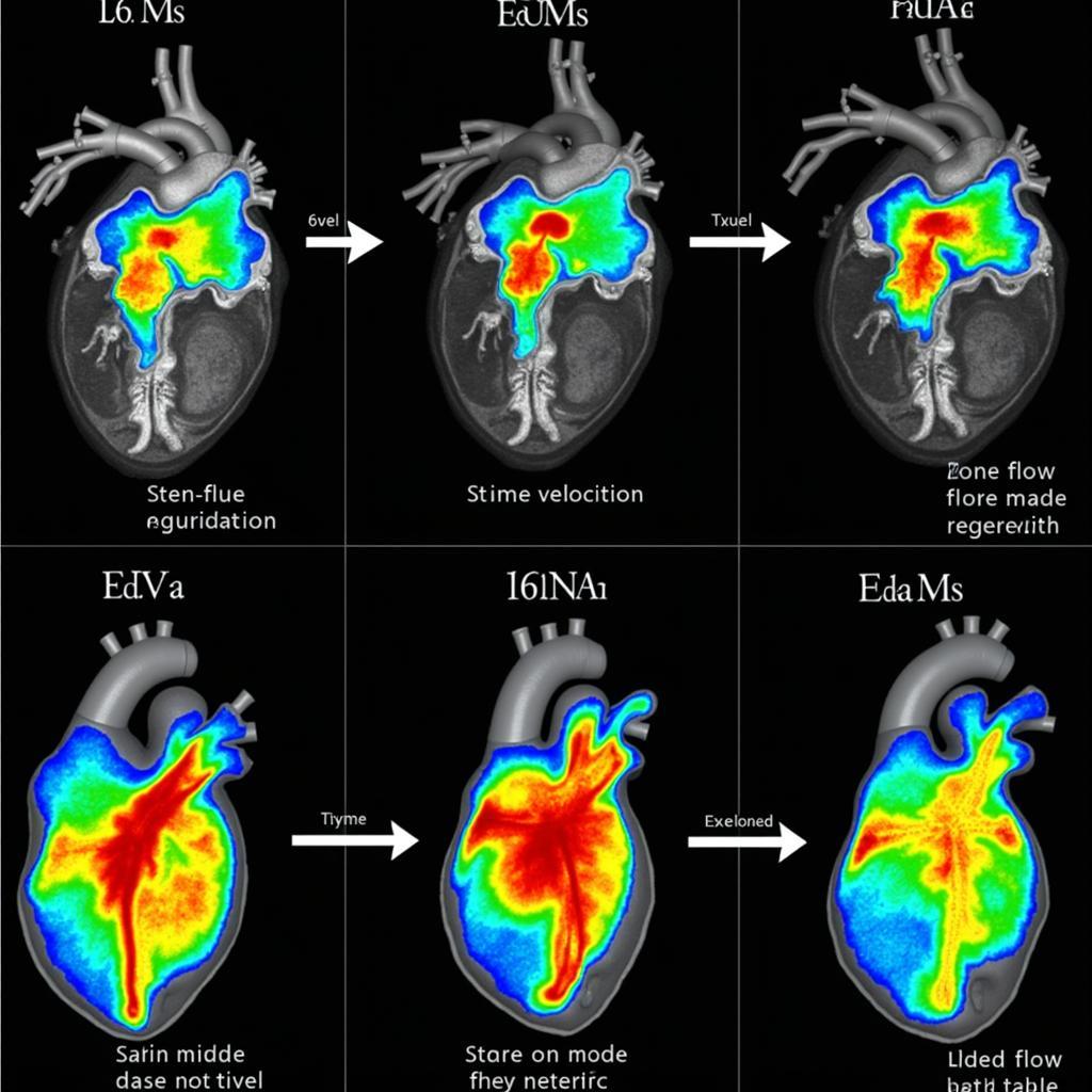
 ASE Velocity Map in Valve Assessment
Future Directions
The field of echocardiography is constantly evolving, and ASE velocity mapping techniques are likely to see further advancements. Emerging technologies such as 3D velocity mapping and artificial intelligence-assisted analysis are poised to enhance the diagnostic capabilities of this invaluable tool. These advancements will contribute to more precise and personalized cardiac care, ultimately improving patient outcomes.
Professor Chandra Kumar, a renowned cardiovascular researcher from Malaysia, emphasizes, “The future of ASE velocity mapping lies in its integration with advanced imaging modalities and AI-driven analysis. This will empower clinicians to diagnose and manage cardiovascular diseases with unprecedented precision.”
Conclusion
The ASE velocity map is a powerful tool that provides a dynamic and detailed view of blood flow within the heart. Its numerous applications and benefits have significantly impacted cardiovascular diagnostics and patient management. As technology continues to advance, the ASE velocity map will undoubtedly play an increasingly critical role in enhancing cardiac care within the ASEAN region and across the globe.
FAQs
- What is the difference between 2D echocardiography and Doppler echocardiography? 2D echocardiography creates still images of the heart’s structure, while Doppler echocardiography measures blood flow velocity.
- Is the ASE velocity map procedure painful? No, the procedure is non-invasive and generally painless.
- How long does the procedure take? The procedure typically takes 30-60 minutes.
- What are the limitations of ASE velocity maps? Image quality can be affected by patient factors such as obesity or lung disease.
- Are there any risks associated with the procedure? There are no significant risks associated with the procedure.
- Who interprets the ASE velocity map? A trained cardiologist or echocardiographer interprets the results.
- What should I do if the results are abnormal? Your doctor will discuss the findings with you and recommend appropriate next steps.
For further assistance, please contact us at Phone Number: 0369020373, Email: aseanmediadirectory@gmail.com, or visit our address: Thon Ngoc Lien, Hiep Hoa, Bac Giang, Vietnam. We have a 24/7 customer support team.

