Ase Normal Fractional Shortening Normal Values are a crucial aspect of assessing cardiac function, particularly left ventricular systolic function. Understanding these values allows medical professionals to identify potential heart issues and provide appropriate treatment. This article delves into the specifics of ASE (American Society of Echocardiography) normal fractional shortening values, their significance, and factors influencing them.
What are ASE Normal Fractional Shortening Values?
Fractional shortening (FS) is a measurement derived from echocardiography that quantifies the percentage change in left ventricular diameter during systole (contraction) and diastole (relaxation). The ASE has established normal reference ranges for FS, typically between 28% and 45%. Values below this range can indicate impaired left ventricular systolic function, potentially signaling conditions like heart failure or cardiomyopathy.
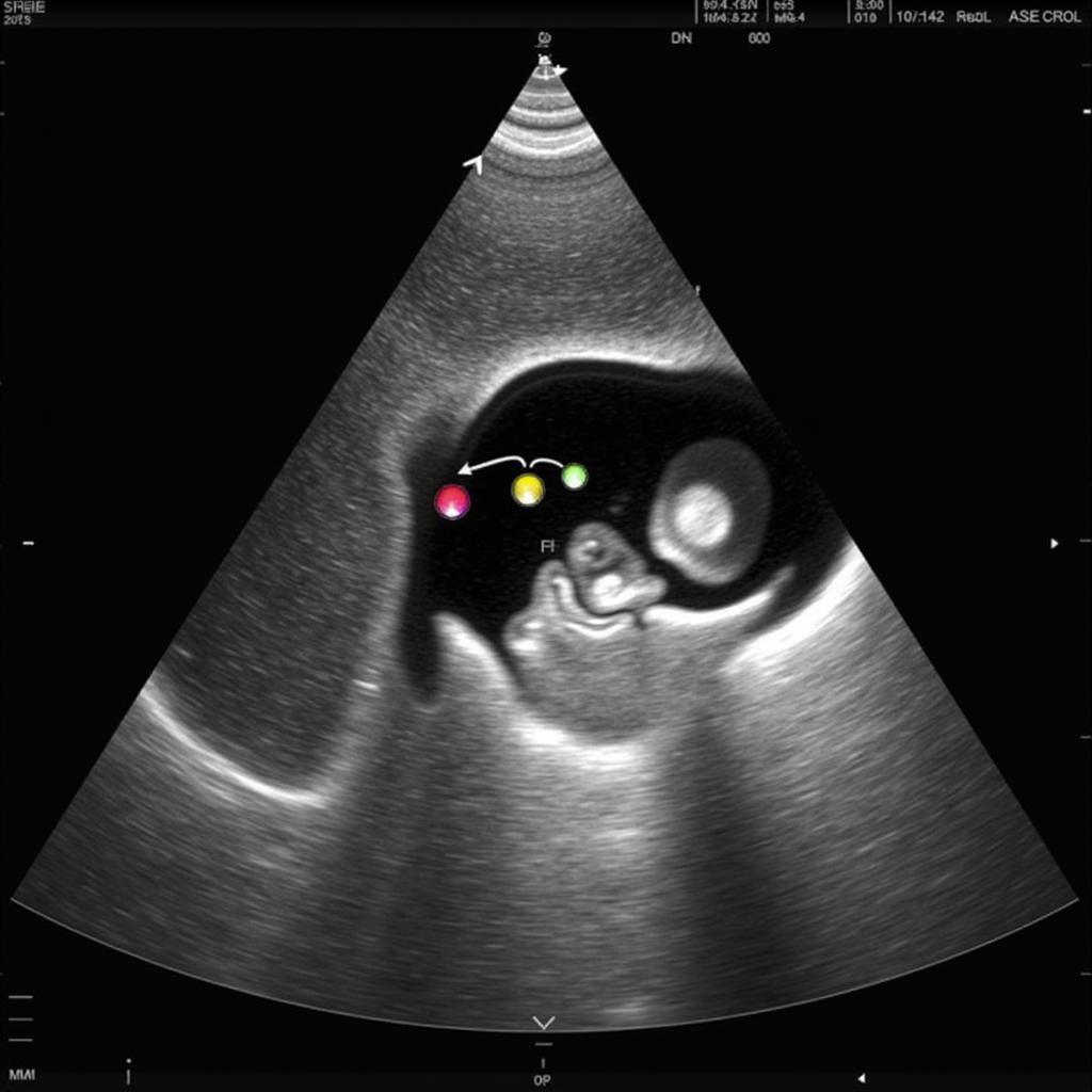
 Echocardiogram showing normal fractional shortening
Why are ASE Normal Fractional Shortening Normal Values Important?
Accurate assessment of FS is critical for early diagnosis and management of cardiovascular diseases. Knowing the ASE normal fractional shortening normal values allows physicians to identify deviations and investigate potential underlying causes. Early detection of abnormalities can significantly impact treatment outcomes and patient prognosis. Moreover, these values provide a baseline for monitoring the effectiveness of therapies and tracking disease progression.
Factors Affecting Fractional Shortening
Several factors can influence FS values, including age, gender, and underlying health conditions. For example, older adults may naturally have slightly lower FS values than younger individuals. Certain medications can also affect heart function and consequently, FS. Therefore, it’s essential to consider these individual factors when interpreting FS measurements.
Interpreting ASE Normal Fractional Shortening Normal Values
While the established range provides a general guideline, interpreting FS requires considering the overall clinical picture. A slightly lower FS value may not always indicate a problem, especially in asymptomatic individuals. However, consistent values below the normal range warrant further investigation.
ASE Guidelines and Strain Imaging
The ASE provides detailed guidelines on performing and interpreting echocardiographic measurements, including FS. These guidelines are regularly updated to reflect the latest advancements in echocardiography. Furthermore, ASE comprehensive strain imaging offers a more sophisticated assessment of myocardial deformation and can provide valuable insights in cases where FS alone may be inconclusive.
ase comprehensive strain imaging
Conclusion
ASE normal fractional shortening normal values are an essential tool for evaluating cardiac function. Understanding these values, their significance, and influencing factors is crucial for healthcare professionals involved in cardiovascular care. By utilizing these guidelines, physicians can accurately assess and manage heart conditions, ultimately improving patient outcomes. Remember, if your FS falls outside the normal range, consult a cardiologist for a comprehensive evaluation.
FAQs
-
What is the normal range for fractional shortening?
The normal range is typically between 28% and 45%. -
What does a low fractional shortening indicate?
It can suggest impaired left ventricular systolic function. -
What factors can affect fractional shortening values?
Age, gender, medications, and underlying health conditions. -
What is the role of ASE in fractional shortening measurement?
The ASE provides guidelines and recommendations for accurate measurement. -
How can I learn more about ASE normal fractional shortening normal values?
Consult a cardiologist or refer to the ASE guidelines. -
What other imaging techniques are used to assess heart function?
Other techniques include MRI, CT scans, and nuclear stress tests. -
What should I do if my fractional shortening is abnormal?
Consult a cardiologist for further evaluation and guidance.
Common Scenarios and Questions
- Scenario: A patient presents with shortness of breath and fatigue. Their FS is found to be low. Question: What are the next steps in their evaluation?
- Scenario: An athlete has a slightly lower FS than average. Question: Is this cause for concern?
- Scenario: A patient’s FS improves after starting medication. Question: How is this improvement interpreted?
Further Reading and Related Articles
- Explore the ASE website for detailed information on echocardiography.
- Search for articles on left ventricular systolic function and heart failure.
- Find resources on cardiovascular health and disease management.
Need Assistance?
For any queries or support regarding cardiovascular health, please contact us:
Phone: 0369020373
Email: [email protected]
Address: Thon Ngoc Lien, Hiep Hoa, Bac Giang, Vietnam
Our customer service team is available 24/7 to assist you.


