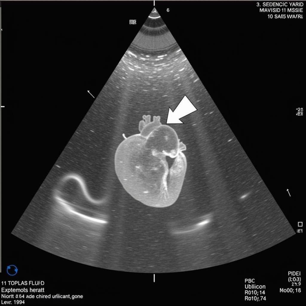
Ase Pediatric Echo Views are crucial for accurately assessing heart function in children. Understanding these standardized views ensures comprehensive evaluations and aids in diagnosing various cardiac conditions. This guide provides a detailed overview of essential ASE pediatric echo views, their significance, and practical applications.
Understanding the Importance of Standardized ASE Pediatric Echo Views
Standardized views in pediatric echocardiography, as recommended by the American Society of Echocardiography (ASE), are essential for consistent and reliable cardiac assessments. These views ensure that all crucial heart structures are visualized systematically, facilitating accurate diagnoses and enabling effective communication between medical professionals. Using ASE guidelines ensures comprehensive examinations and reduces the risk of overlooking subtle abnormalities.  ASE Pediatric Echo Parasternal Long Axis View
Consistent application of ASE pediatric echo views also allows for accurate comparisons across different examinations, monitoring disease progression, and evaluating treatment effectiveness. This standardization is particularly vital in pediatric cardiology, where early and precise diagnosis is critical for optimal patient outcomes.
Key ASE Pediatric Echo Views and Their Applications
Several key ASE pediatric echo views form the cornerstone of comprehensive cardiac assessments. These include:
- Parasternal Long Axis View: This view visualizes the left ventricle, left atrium, mitral valve, aortic valve, and right ventricular outflow tract. It’s essential for assessing ventricular function, valve morphology, and the relationship between these structures.
- Parasternal Short Axis View: Obtained at various levels, this view provides cross-sectional images of the ventricles, atria, and valves. It’s critical for evaluating ventricular wall thickness, chamber size, and valve function.
- Apical Four-Chamber View: This view displays all four chambers of the heart simultaneously, allowing assessment of chamber size, atrial and ventricular septal integrity, and valve function. It’s crucial for identifying congenital heart defects.
- Subcostal View: This view, obtained with the transducer placed below the rib cage, is particularly useful in infants and young children. It provides excellent visualization of the inferior vena cava, hepatic veins, and atrial septum.
Utilizing ASE Pediatric Echo Views in Specific Congenital Heart Defects
ASE pediatric echo views play a pivotal role in diagnosing and managing various congenital heart defects. For example, in Tetralogy of Fallot, the parasternal short axis view is crucial for visualizing the overriding aorta and ventricular septal defect. Similarly, the apical four-chamber view is essential for assessing atrial and ventricular septal defects in patients with Atrioventricular Septal Defect (AVSD). ase standard echo protocol
What are the common challenges in obtaining optimal ASE pediatric echo views?
Obtaining optimal ASE pediatric echo views can be challenging, especially in young or uncooperative patients. Respiratory variations, patient movement, and body habitus can affect image quality. Experienced sonographers employ various techniques, including patient positioning, distraction methods, and adjusting transducer frequency and settings, to overcome these challenges. ase pediatric echo guidelines
How can image quality be optimized during pediatric echocardiography?
Optimizing image quality in pediatric echocardiography involves adjusting factors like transducer frequency, depth, and gain settings. Using harmonics and compound imaging can improve image resolution and reduce artifacts. Furthermore, proper patient positioning and minimizing patient movement are crucial for obtaining clear and diagnostic images.
Conclusion
ASE pediatric echo views are fundamental to pediatric cardiology. These standardized views ensure comprehensive and reliable cardiac assessments, enabling accurate diagnosis and effective management of congenital and acquired heart diseases. Adhering to ASE guidelines is essential for all medical professionals performing pediatric echocardiography. Understanding and consistently applying these views contributes significantly to improved patient care and outcomes in pediatric cardiology. Applying these ASE pediatric echo views correctly leads to better diagnostic accuracy and treatment planning. ase exam fetal echocardiography
FAQ
- What is the purpose of ASE pediatric echo views?
- What are the key ASE pediatric echo views?
- How are ASE pediatric echo views used in diagnosing congenital heart defects?
- What challenges can arise when obtaining ASE pediatric echo views?
- How can image quality be optimized in pediatric echocardiography?
- Why is standardization important in pediatric echocardiography?
- Where can I find more information on ASE guidelines for pediatric echocardiography?
Scenarios
- Scenario 1: A newborn presents with cyanosis. ASE pediatric echo views are used to diagnose Tetralogy of Fallot.
- Scenario 2: A child with a heart murmur undergoes echocardiography. ASE views are used to evaluate valve function and diagnose mitral valve prolapse.
- Scenario 3: A fetus is suspected of having a congenital heart defect. Fetal echocardiography using ASE views confirms the diagnosis. ase fetal echo guidelines
Further Reading
Explore related articles on ase posters for more in-depth information.
When you need assistance, please contact Phone Number: 0369020373, Email: [email protected] Or visit us at: Thôn Ngọc Liễn, Hiệp Hòa, Bắc Giang, Việt Nam. We have a 24/7 customer service team.

