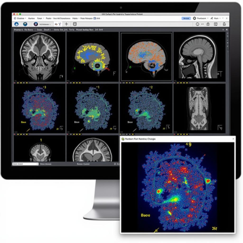
Strain rate imaging, a sophisticated echocardiographic technique, plays a crucial role in evaluating cardiac function, particularly myocardial deformation. This technique is guided by the meticulous standards outlined by the American Society of Echocardiography (ASE), ensuring accuracy and consistency in clinical practice. This article delves into the importance of ASE guidelines for strain rate imaging, highlighting their impact on diagnosis and patient care.
 ASE Guidelines for Strain Rate Imaging
The Significance of Standardized Strain Rate Imaging
Strain rate imaging provides invaluable insights into the mechanics of the heart, allowing cardiologists to assess how well the heart muscle contracts and relaxes. However, the complexity of this technique necessitates standardized procedures to minimize variability and ensure reliable results. This is where ASE guidelines come into play.
These guidelines, developed by experts in the field, provide a framework for:
- Image Acquisition: Specifying optimal patient positioning, transducer selection, and imaging planes to obtain high-quality images.
- Measurement Techniques: Outlining precise methods for tracing myocardial contours and measuring strain rate values, minimizing operator dependence.
- Interpretation of Results: Establishing normal reference values and criteria for identifying abnormal myocardial function based on strain rate parameters.
 Analyzing Strain Rate Imaging Results
Key Aspects of ASE Guidelines for Strain Rate Imaging
The ASE guidelines address several key aspects of strain rate imaging:
- Modalities: They provide recommendations for different strain rate imaging modalities, including 2D speckle tracking echocardiography (2D STE) and tissue Doppler imaging (TDI).
- Clinical Applications: The guidelines detail the clinical scenarios where strain rate imaging is particularly valuable, such as in the evaluation of coronary artery disease, heart failure, and valvular heart disease.
- Technical Considerations: They highlight important technical factors that can influence the accuracy of strain rate measurements, including image quality, frame rate, and artifacts.
Benefits of Adhering to ASE Guidelines
Adherence to ASE guidelines for strain rate imaging offers several benefits:
- Enhanced Accuracy: Standardized protocols reduce variability, leading to more accurate and reliable measurements of myocardial strain rate.
- Improved Reproducibility: Guidelines ensure consistency in image acquisition and analysis, enabling different clinicians to obtain comparable results.
- Enhanced Diagnostic Confidence: By providing standardized criteria for interpretation, the guidelines increase confidence in diagnosing and monitoring cardiac conditions.
- Facilitated Research: Standardized methods enable meaningful comparisons of research findings across different studies and institutions.
ASE Guidelines: A Cornerstone for Cardiac Imaging
The ASE guidelines for strain rate imaging represent a cornerstone of quality assurance in cardiac imaging. They empower cardiologists to leverage the full potential of this powerful technique for accurate diagnosis, personalized treatment plans, and ultimately, improved patient outcomes. By adhering to these guidelines, healthcare professionals contribute to a higher standard of care in cardiology, harnessing the power of precision medicine for the benefit of patients worldwide.
