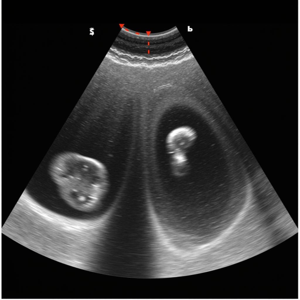
Cardiomyopathy, a complex group of diseases affecting the heart muscle, poses significant diagnostic and management challenges. The American Society of Echocardiography (ASE) guidelines for cardiomyopathy, often referred to as “Ase Guidelines Cardiomyopathy,” provide crucial recommendations for utilizing echocardiography in the evaluation and care of patients with this condition. These guidelines offer valuable insights into various aspects, from image acquisition and interpretation to disease classification and prognostication.
 ASE Guidelines for Cardiomyopathy Diagnosis
Types of Cardiomyopathy and ASE Recommendations
The ASE guidelines address various types of cardiomyopathy, including dilated cardiomyopathy (DCM), hypertrophic cardiomyopathy (HCM), restrictive cardiomyopathy (RCM), and arrhythmogenic right ventricular cardiomyopathy (ARVC). Each type presents unique echocardiographic characteristics, and the guidelines offer specific recommendations for their assessment. For instance, in DCM, the ASE emphasizes the importance of assessing left ventricular size and function, while in HCM, the focus is on identifying left ventricular hypertrophy and assessing left ventricular outflow tract obstruction. ase chamber quantification 2017 provides further details on chamber quantification techniques.
Dilated Cardiomyopathy (DCM)
The ASE guidelines for DCM emphasize meticulous assessment of left ventricular size and systolic function using various echocardiographic parameters. These include left ventricular ejection fraction (LVEF), fractional shortening, and left ventricular end-diastolic and end-systolic dimensions.
Hypertrophic Cardiomyopathy (HCM)
In HCM, the guidelines highlight the crucial role of echocardiography in identifying left ventricular hypertrophy, assessing its distribution, and evaluating the presence and severity of left ventricular outflow tract obstruction.
 ASE Guidelines for HCM LVOT Assessment
The Importance of Strain Imaging
Strain imaging, a sophisticated echocardiographic technique, plays an increasingly important role in the evaluation of cardiomyopathy. The ASE guidelines recognize the value of strain imaging in assessing myocardial deformation and identifying subtle abnormalities in myocardial function, even in patients with preserved LVEF. This technique can provide valuable prognostic information and help guide treatment decisions. “Strain imaging allows us to detect early signs of myocardial dysfunction, which can be crucial for timely intervention,” says Dr. Emily Carter, a leading cardiologist specializing in cardiomyopathy.
Utilizing Strain Imaging in Cardiomyopathy
The ASE guidelines provide detailed recommendations on how to perform and interpret strain imaging in different types of cardiomyopathy. They emphasize the importance of standardized protocols and quality control to ensure accurate and reproducible results.
ASE Guidelines and Pericardial Disease
While the primary focus of the “ase guidelines cardiomyopathy” is on myocardial diseases, the guidelines also address the echocardiographic assessment of pericardial diseases, which can sometimes mimic or coexist with cardiomyopathy. ase guidelines pericardial disease offers further information on this topic. Understanding these conditions is crucial for accurate diagnosis and management.
“Accurate assessment of pericardial disease is essential to rule out or confirm its contribution to a patient’s symptoms,” adds Dr. David Lee, a renowned expert in cardiac imaging.
Conclusion
The ASE guidelines for cardiomyopathy provide a comprehensive framework for utilizing echocardiography in the evaluation and management of patients with this complex group of diseases. By adhering to these guidelines, clinicians can ensure accurate diagnosis, appropriate risk stratification, and optimal patient care. Understanding “ase guidelines cardiomyopathy” is key for any healthcare professional involved in the care of these patients. ase mitral regurg can offer additional insights into mitral regurgitation, a common complication of cardiomyopathy. Remember that staying updated with the latest ASE guidelines is crucial for providing the best possible care. ase using hall health is a good resource for continued learning.
FAQ
- What are the main types of cardiomyopathy?
- How does echocardiography help in diagnosing cardiomyopathy?
- What is strain imaging, and why is it important in cardiomyopathy?
- How do the ASE guidelines address pericardial disease?
- Where can I find the latest ASE guidelines for cardiomyopathy?
- What is the role of ASE chamber quantification in evaluating cardiomyopathy?
- How does mitral regurgitation relate to cardiomyopathy?
Common Scenarios and Questions:
- Scenario: A patient presents with shortness of breath and fatigue. Question: How can echocardiography differentiate between different types of cardiomyopathy?
- Scenario: A patient with HCM experiences chest pain. Question: How can echocardiography assess the severity of LVOT obstruction?
- Scenario: A patient with DCM has preserved LVEF. Question: How can strain imaging help evaluate myocardial function?
Further Exploration:
You might also be interested in learning more about asean agreements.
Contact Us
For any assistance, please contact us at Phone Number: 0369020373, Email: aseanmediadirectory@gmail.com or visit us at Thôn Ngọc Liễn, Hiệp Hòa, Bắc Giang, Việt Nam. We have a 24/7 customer service team.
